Epicondyle Of Femur, Medial Femoral Condyle Anatomyzone
Epicondyle of femur Indeed recently is being sought by users around us, maybe one of you. Individuals now are accustomed to using the net in gadgets to see image and video data for inspiration, and according to the title of the article I will discuss about Epicondyle Of Femur.
- Human Femur Bones Royalty Free Vector Image Vectorstock
- Medial Epicondyle Of The Femur Wikiwand
- Condyle Transparent Background Png Cliparts Free Download Hiclipart
- Femoral Shaft Fractures Core Em
- Resolve Your Knee Pain By Addressing Its Alignment Msk Neurology
- Skill Lab Learning
Find, Read, And Discover Epicondyle Of Femur, Such Us:
- Medial Epicondyle Of The Femur Wikipedia
- Femur Bone Anatomy Proximal Distal And Shaft Kenhub
- Femur Definition Function Diagram Facts Britannica
- Medial Epicondyle High Resolution Stock Photography And Images Alamy
- Distal Femur Fractures Trauma Orthobullets
If you are looking for Endocrinologist Near Me For Adults you've come to the right location. We have 104 images about endocrinologist near me for adults including images, photos, pictures, backgrounds, and much more. In such page, we also provide number of images available. Such as png, jpg, animated gifs, pic art, logo, black and white, translucent, etc.
Crossfit Bones Of The Knee Endocrinologist Near Me For Adults
Anatomy Knee Endocrinologist Near Me For Adults

The Photo Shows The Attachments On The Lateral Epicondyle Of Femur And Download Scientific Diagram Endocrinologist Near Me For Adults
Bones Advanced Anatomy 2nd Ed Endocrinologist Near Me For Adults
Condyle Transparent Background Png Cliparts Free Download Hiclipart Endocrinologist Near Me For Adults
What Is The Difference Between Condyle And Epicondyle Quora Endocrinologist Near Me For Adults
52 of hip fractures occur after the age of 80 years.
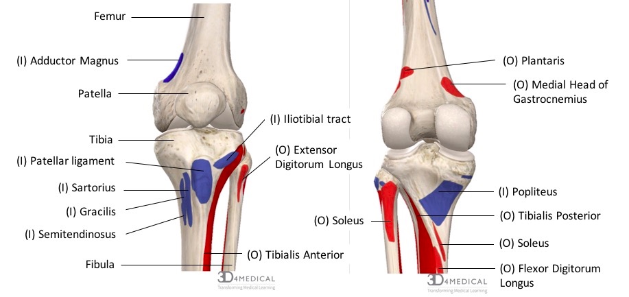
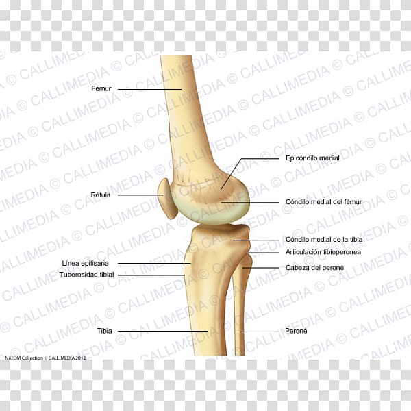
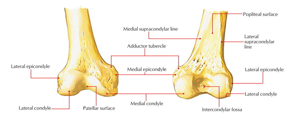
Endocrinologist near me for adults. Prominent lateral and medial condyles are found at the distal end of the femur. Directly below it is a small depression from which a smooth well marked groove curves obliquely upward and backward to the posterior extremity of the condyle. The iliopatellar band.
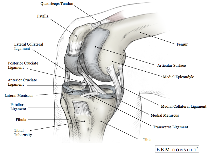
This page was last edited on 27. The medial and lateral collateral ligaments of the knee originate from their respective epicondyles. Projecting from each condyle is an epicondyle that act as attachment sites for the collateral ligaments.
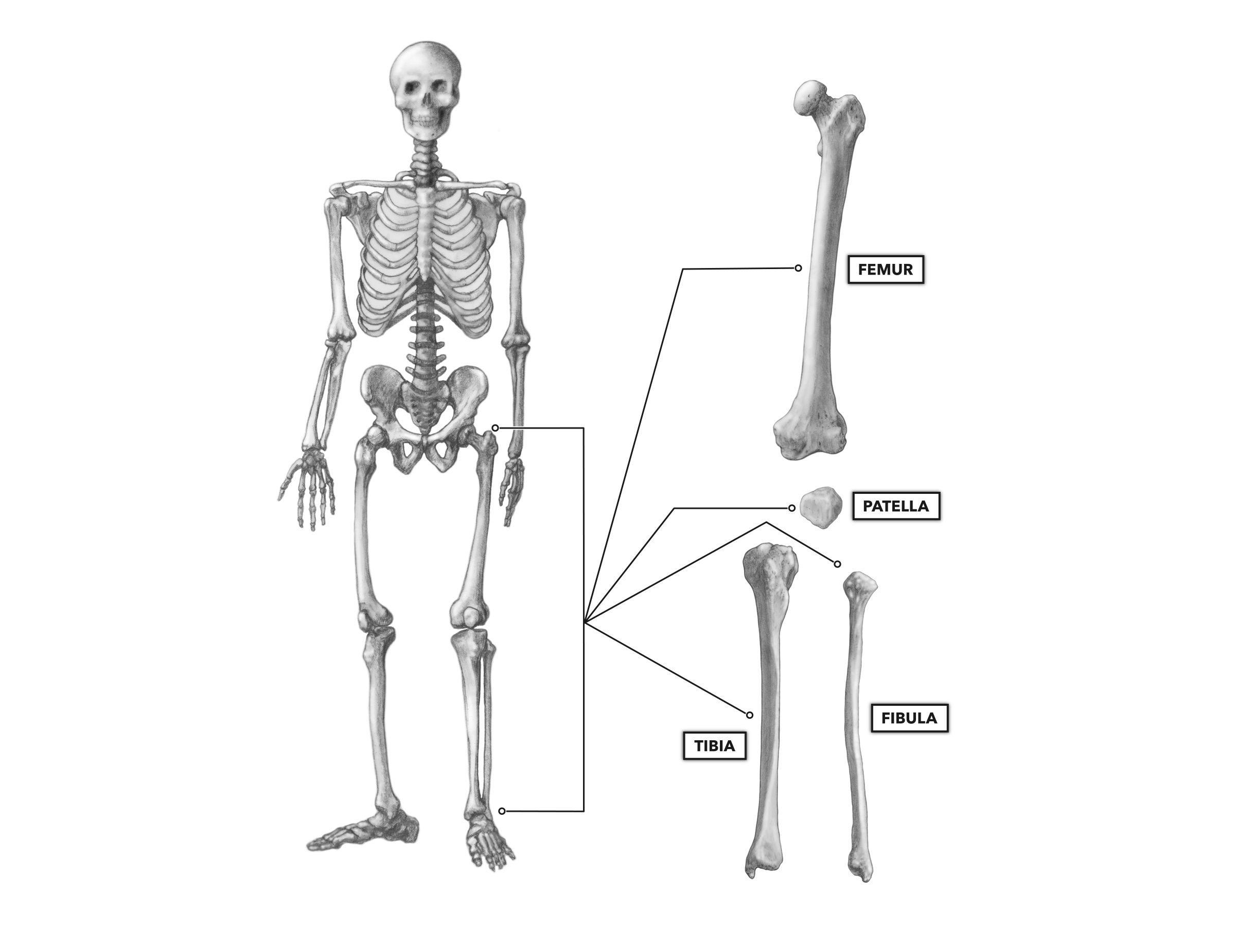
The femur tibia and patella. The iliotibial tract continues downward laterally from the femur. The lateral epicondyle of the femur smaller and less prominent than the medial epicondyle gives attachment to the fibular collateral ligament of the knee joint.
Lateral epicondyle of the femur this human musculoskeletal system article is a stub. Femoral shaft diaphysis. You can help wikipedia by expanding it.
Radius and ulna shaft diaphysis monteggia fracture dislocation. It contains two facets for attachment of intracapsular knee ligaments. The knee also known as the tibiofemoral joint is a synovial hinge joint formed between three bones.
Two rounded convex processes known as condyles on the distal end of the femur meet two rounded concave condyles at the proximal end of the tibia. Wrist distal radius and ulna. X worldwide proximal femoral neck fracture more commonly known as hip fracture is a frequent serious injury of older people 1 and increases the risk of death and major morbidity.
Lateral epicondyle of the humerus dorsal epicondyle in birds in humans on the outboard side of the elbow. Physeal growth plate hand hip and proximal femur. If there is a fracture break in part of the condyle this is known as a fracture of the femoral condyle.
The lateral and medial condyles are separated by the intercondylar notch. When approaching the knee joint the iliotibial tract passes the lateral epicondyle of the femur and splits into two structures. A femoral condyle is the ball shape located at the end of the femur thigh bone.
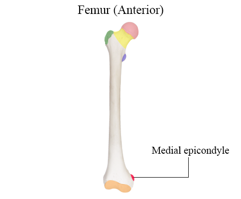
Intercondylar fossa a deep notch on the posterior surface of the femur between the two condyles. Medial epicondyle of the femur. The femoral artery is the main blood supply to the lower.
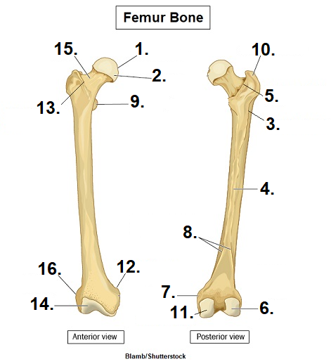
There are two condyles on each leg known as the medial and lateral femoral condyles. At the greater trochanter fibres of the tensor fascia lata muscle and gluteus maximus muscle inserts in the iliotibial tract. The femur bone is the strongest and longest bone in the body occupying the space of the lower limb between the hip and knee jointsfemur anatomy is so unique that it makes the bone suitable for supporting the numerous muscular and ligamentous attachments within this region in addition to maximally extending the limb during ambulation.
Easy Notes On Femur Learn In Just 4 Minutes Earth S Lab Endocrinologist Near Me For Adults

The Knee Musculoskeletal Key Endocrinologist Near Me For Adults

Knee Joint D Rania Gabr D Sama D Elsherbiny Ppt Video Online Download Endocrinologist Near Me For Adults
Skill Lab Learning Endocrinologist Near Me For Adults
More From Endocrinologist Near Me For Adults
- Thoracic Spine Anatomy Numbers
- Hinge Joints Concrete
- Diagram Hinge Joint Examples
- Hinge Joint Hrvatski
- Unity Hinge Joint Child
Incoming Search Terms:
- Screenshot Of Distal Femur Including Roi Points For Sulcus Line Medial Download Scientific Diagram Unity Hinge Joint Child,
- Crossfit Bones Of The Knee Unity Hinge Joint Child,
- Pomf Week 2 Knee Phyt606 Aut Pomf Week The Knee Joint Bony Landmarks Of Studocu Unity Hinge Joint Child,
- Anatomy Of The Knee Joint Paley Orthopedic Spine Institute Unity Hinge Joint Child,
- The Femur Pelvic Girdle Unity Hinge Joint Child,
- Condyle Transparent Background Png Cliparts Free Download Hiclipart Unity Hinge Joint Child,
