Lateral Epicondyle Of Femur Palpation, Facebook
Lateral epicondyle of femur palpation Indeed recently has been hunted by consumers around us, maybe one of you personally. People are now accustomed to using the internet in gadgets to view video and image information for inspiration, and according to the name of this article I will talk about about Lateral Epicondyle Of Femur Palpation.
- Collateral Ligament Injuries Of The Knee Orthopaedia
- Contents Preface Acknowledgments List Of Clinical Blue Boxes List Of Tables Figure Credits 1 Overview And Basic Concepts Approaches To Studying Anatomy Regional Anatomy Systemic Anatomy Clinical Anatomy Anatomicomedical Terminology
- Palpation Guided Anatomic Markers Anatomic Landmarks Are First Download Scientific Diagram
- Medial Knee Injuries Wikipedia
- Physeal Sparing Medial Patellofemoral Ligament Reconstruction With Suture Anchor For Femoral Graft Fixation Arthroscopy Techniques
- The Lower Limb Basicmedical Key
Find, Read, And Discover Lateral Epicondyle Of Femur Palpation, Such Us:
- The Lower Limb Basicmedical Key
- Lateral Epicondylitis Teachmesurgery
- Palpation Guided Anatomic Markers Anatomic Landmarks Are First Download Scientific Diagram
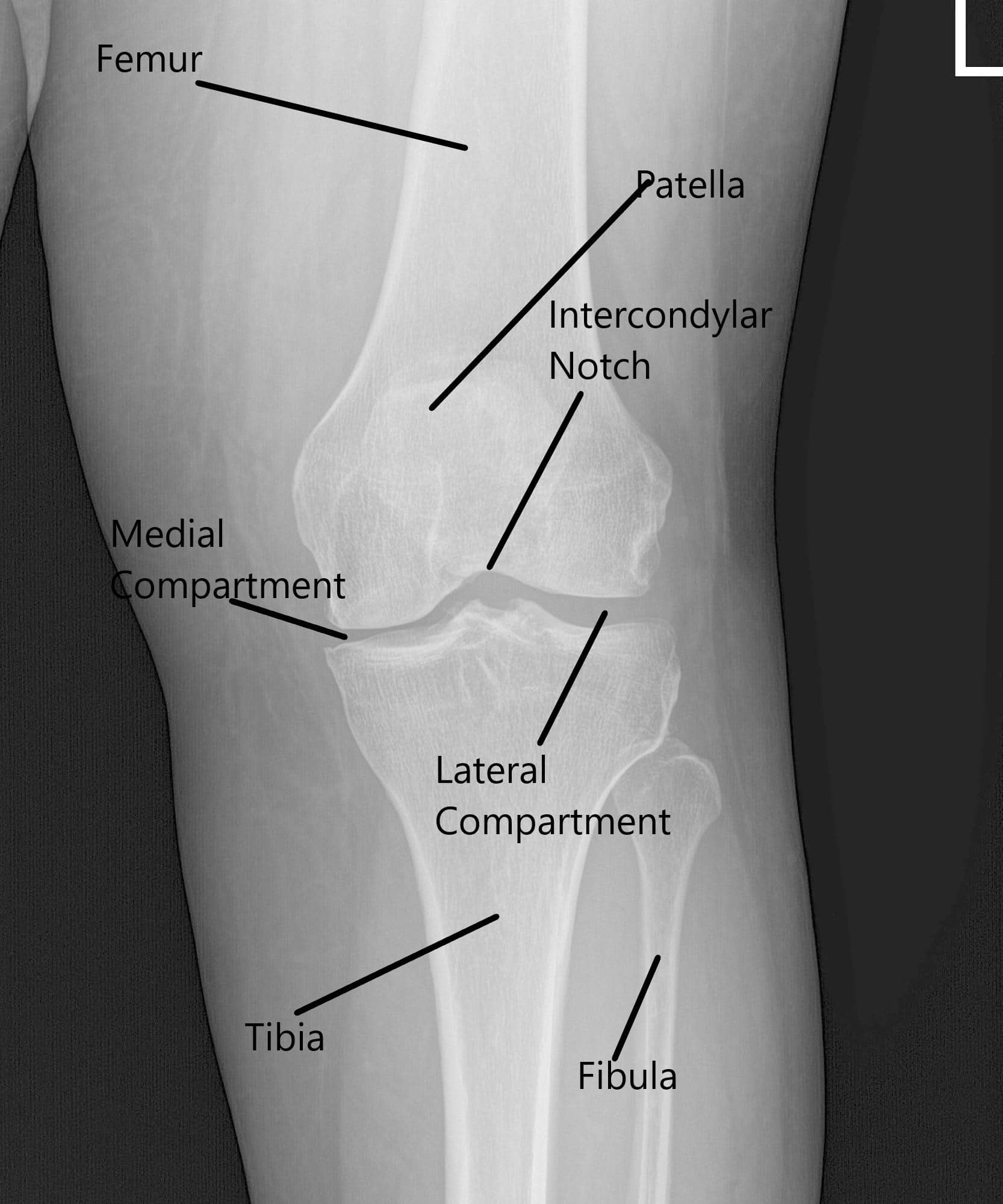
- Knee Anatomy Laurasloan2
- Athletic Training Sports Medicine Center Home Athletic Training Sports Medicine Center Ah 321 Assessment Of Athletic Injuries Illnesses Laboratory Bony Palpation Skill Sheet Hand Wrist Arm Hip Pelvis Spine Foot Ankle Forearm Shoulder
If you re searching for Hinge Offset Joint Kafo you've arrived at the ideal location. We have 104 images about hinge offset joint kafo adding images, photos, photographs, backgrounds, and much more. In these web page, we additionally provide variety of graphics out there. Such as png, jpg, animated gifs, pic art, symbol, black and white, translucent, etc.

Medial Knee Injuries Wikipedia Hinge Offset Joint Kafo
Case Study Medial Meniscus Root Repair With Endobutton And Chondroplasty Of The Left Knee Hinge Offset Joint Kafo
Knee Injuries Hinge Offset Joint Kafo

Femur Posterolateral Approach Approaches Orthobullets Hinge Offset Joint Kafo

Supine Right Knee Marks Are Made On The Knee After Percutaneous Download Scientific Diagram Hinge Offset Joint Kafo

Palpation Antero Lateral Aspect 13 Knee Joint Anatomy Of The Knee Joint Hinge Offset Joint Kafo
Pelvis femur trauma.
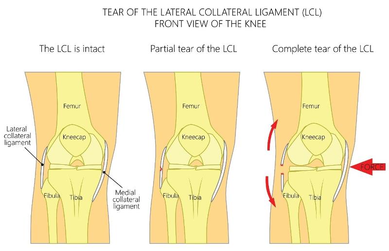
Hinge offset joint kafo. Netters atlas of human anatomy 5th edition. Ecchymosis and lateral joint soft tissue swelling. Runs between the lateral epicondyle of the femur and head of the fibula.
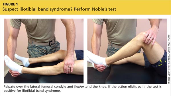
It usually starts with lateral knee pain during and after runs but there are two major types. Paul ingraham updated jan 5 2019 runners knee refers to one of two common1 repetitive strain injuries of the knee either iliotibial band syndrome lateral knee pain or patellofemoral syndrome anterior knee pain. However the definition in human anatomy refers only to the section of the lower limb extending from the knee to the ankle also known as the crus or especially in non technical use the shank.
Both usually affect runners triathletes. Lateral collateral ligament lcl prevents medial movement of the tibia on the femur when varus towards the midline stress is placed on the knee. He has exquisite tenderness to palpation along the lateral aspect of his elbow.
Netters atlas of human anatomy 5th edition download. Lateral epicondyle of the femur. The human leg in the general word sense is the entire lower limb of the human body including the foot thigh and even the hip or gluteal region.
At the proximal level this ligament is closely related to the joint capsule without having direct contact as it is separated by fat pad the insertion is augmented by the iliotibial band. Menisci the medial and lateral menisci are located within the knee joint attached to the tibial plateau. Some authors describe the lateral patellofemoral ligament as a palpable thickening of the joint capsule between the patella and femoral epicondyle.
Patellofemoral pain syndrome pfps is the most common cause of knee pain in the outpatient setting. It is caused by imbalances in the forces controlling patellar tracking during knee flexion and. The lcl further splits the biceps femoris into two parts.
Entire length of ligament can be palpated by placing patient in figure of 4 position. The lateral patellofemoral ligament lpfl is an important lateral stabilizer of the patella against medial subluxation or dislocation. Hyperextension or varus lateral.
Legs are used for standing and all forms of. Together they fuse to the distal humerus between the ages of 14 16 years old. Intact ligament will be a palpable cordlike structure.
Tenderness over lcl insertion. Lateral joint line pain and swelling. What additional radiographic view will.
Nontraumatic Knee Pain A Diagnostic Treatment Guide Clinician Reviews Hinge Offset Joint Kafo
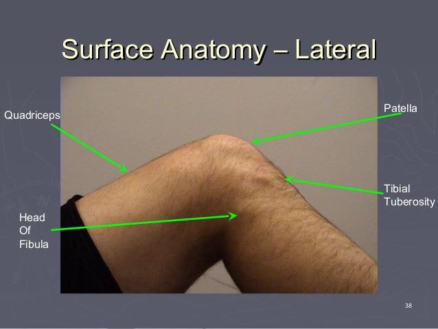
Surface Anatomy Hinge Offset Joint Kafo

Femoral Condyle Osteochondral Fracture Treated With Bone Suture After Acute Patellar Dislocation A Case Report Hinge Offset Joint Kafo

Collateral Ligament Injuries Of The Knee Orthopaedia Hinge Offset Joint Kafo
More From Hinge Offset Joint Kafo
- Hinge Joints Tagalog
- Diseases Of The Endocrine System In Animals
- Hinge Joint Field Fence
- Spine Definition Anatomy Quizlet
- Lower Back Muscle Pain Anatomy
Incoming Search Terms:
- The Bone Attachments Of The Medial Collateral And Posterior Oblique Ligaments Are Defined Anatomically And Radiographically Springerlink Lower Back Muscle Pain Anatomy,
- Ppt Knee Lab Powerpoint Presentation Free Download Id 160856 Lower Back Muscle Pain Anatomy,
- Http Eprints Usm My 39817 1 Dr Mohd Saiful Adzuwan Mat Rodi 24 Pages Pdf Lower Back Muscle Pain Anatomy,
- Disorders Of Lower Limb Lower Back Muscle Pain Anatomy,
- Anatomy Of The Canine Knee Easyanatomy Lower Back Muscle Pain Anatomy,
- Ppt Femur Patellar Surface Femur Lateral Condyle Epicondyle Head Fibula Tibial Tuberosity Fibula Powerpoint Presentation Id 3340347 Lower Back Muscle Pain Anatomy,
