Lateral Epicondyle Of Femur Function, Pomf Week 2 Knee Phyt606 Aut Pomf Week The Knee Joint Bony Landmarks Of Studocu
Lateral epicondyle of femur function Indeed recently is being sought by consumers around us, maybe one of you. Individuals are now accustomed to using the net in gadgets to view image and video data for inspiration, and according to the name of this article I will talk about about Lateral Epicondyle Of Femur Function.
- Thigh Knee And Popliteal Fossa Amboss
- Femoral Shaft Fractures Core Em
- Premium Vector Anatomy Of Human Knee Sketch Of Leg Bones And Joint Medicine Side And Front View Of Knee Bones Hand Drawn Femur Patella Tibia And Fibula Tibial Plateau And
- Figure 1 Two Cases Of Inferior Dislocation Of The Patella With Impaction Into The Femoral Trochlea Of Osteophytes On The Superior Pole Of The Patella
- Anatomy Musculoskeletal Key
- What Is Iliotibial Band Friction Syndrome And What Are Its Causes
Find, Read, And Discover Lateral Epicondyle Of Femur Function, Such Us:
- Native Knee Kinematics Hip Knee Book
- Knee Clinical Gate
- Distal Femoral Fractures Musculoskeletal Key
- Unicondylar Fractures Of The Distal Femur Sciencedirect
- Femur Wikipedia
If you re searching for Immovable Joints In The Body you've reached the ideal place. We ve got 104 graphics about immovable joints in the body including images, photos, photographs, wallpapers, and much more. In such webpage, we also have number of images out there. Such as png, jpg, animated gifs, pic art, symbol, black and white, transparent, etc.

Motion Of The Femoral Condyles In Flexion And Extension During A Continuous Lunge Feng 2015 Journal Of Orthopaedic Research Wiley Online Library Immovable Joints In The Body
Knee Clinical Gate Immovable Joints In The Body

The Iliotibial Band And Site Of Injury At Lateral Epicondyle Of The Femur Download Scientific Diagram Immovable Joints In The Body
Femur Bone Anatomy Proximal Distal And Shaft Kenhub Immovable Joints In The Body

Unicondylar Fractures Of The Distal Femur Sciencedirect Immovable Joints In The Body
Https Encrypted Tbn0 Gstatic Com Images Q Tbn And9gcss Hzjjyfzv U1dmrtheh2xxhzrhbwq4fnot1dzxxrggs6jchq Usqp Cau Immovable Joints In The Body
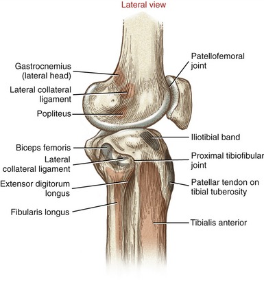
The primary function of the lcl is to protect the knee from varus producing forces figure 10 11 c.

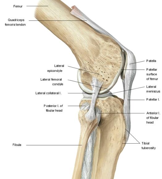
Immovable joints in the body. The femoral artery is the main blood supply to the lower. Likewise the lateral collateral ligament lcl binds the lateral side of the femur to the fibula and prevents forces applied to the medial side of the knee from moving the knee laterally. The lateral and medial condyles are separated by the intercondylar notch.
Medial and lateral epicondyles bony elevations on the non articular areas of the condyles. It is separate from both the joint capsule and the lateral meniscus. The popliteus tendon is deep to the lcl seperating it from the lateral meniscus.
Prominent lateral and medial condyles are found at the distal end of the femur. The lateral condyle also has a shallow groove below the lateral epicondyle through which the popliteal tendon travels. It is caused by repeated use of the superficial extensor muscles which strains their common tendinous attachment to the lateral epicondyle.
It is known as the groove for popliteus. The medial and lateral collateral ligaments of the knee originate from their respective epicondyles. The fibular collateral ligament lcl aka.
The lateral collateral ligament lcl is a round cord like ligament that crosses the lateral side of the knee attaching to the lateral epicondyle of the femur and the head of the fibula. The femur f i m er pl. Fibular stretches from the lateral epicondyle of the femur to the head of fibula.
Long longitudinal axes and expanded ends ex humerus tibia femur. The lcl further splits the biceps femoris into two parts. Intercondylar fossa a deep notch on the posterior surface of the femur between the two condyles.
Lateral epicondyle of the femur. The peak age of onset is 40 50 years old. Lateral epicondylitis or tennis elbow refers to inflammation of the periosteum of the lateral epicondyle.
Two internal ligaments the anterior and posterior cruciate ligaments also help to maintain the proper alignment of the knee. Femurs or femora f m er e or thigh bone is the proximal bone of the hindlimb in tetrapod vertebrate the largest bone of the human bodythe head of the femur articulates with the acetabulum in the pelvic bone forming the hip joint while the distal part of the femur articulates with the tibia and kneecap forming the knee joint. At the proximal level this ligament is closely related to the joint capsule without having direct contact as it is separated by fat pad the insertion is augmented by the iliotibial band.
The medial epicondyle is the larger. The anterolateral ligament all is situated in front of the lcl.
Knee Biomechanics Recon Orthobullets Immovable Joints In The Body

Medial Epicondyle Femur Google Search Skeletal System Muscle Joint Immovable Joints In The Body

Distal Femoral Fractures Musculoskeletal Key Immovable Joints In The Body
Https Www Worldscientific Com Doi Pdf 10 1142 9789814282048 0001 Immovable Joints In The Body
More From Immovable Joints In The Body
- Hinge Joint Wire Spinner
- Human Nervous System Parts And Functions
- Hinge Joint Of Mandible
- Animated Hinge Joint Gif
- Parts Of The Nervous System For Kids
Incoming Search Terms:
- About The Knee Bromsgrove Private Clinic Parts Of The Nervous System For Kids,
- Unicondylar Fractures Of The Distal Femur Sciencedirect Parts Of The Nervous System For Kids,
- Slideshow Femur Parts Of The Nervous System For Kids,
- Osteonecrosis Of The Knee Orthoinfo Aaos Parts Of The Nervous System For Kids,
- Thigh Knee And Popliteal Fossa Amboss Parts Of The Nervous System For Kids,
- Easy Notes On Lateral Condyle Learn In Just 4 Minutes Earth S Lab Parts Of The Nervous System For Kids,
